Bookmark and Share
Showing posts with label Human Disease. Show all posts

Risk Factors for Cancer

Many genetic and environmental factors increase the risk of developing cancer. However, not all people who are exposed to carcinogens or who have other risk factors develop cancer.

Genes That Cause Cancer:

Abnormalities (mutations) affecting critical genes are believed to contribute to the development of cancer. These genes produce proteins that regulate growth and alter cell division and other basic cell properties. Gene mutations causing cancer may result from the damaging effects of chemicals, sunlight, drugs, viruses, or other environmental agents. In some families, these abnormal cancer-causing genes are inherited.

The two major categories of genes involved with cancer are oncogenes and tumor suppressor genes.

Oncogenes are mutated forms of genes that in their normal state regulate cell growth. If they become overactive and signal cells to divide when these cells should not, cancer may develop. The mutation of oncogenes is not entirely understood, but many factors may contribute, including x-rays, sunlight, toxins at work, in the air, or in chemicals (for example, in tobacco smoke), and infectious agents (for example, certain viruses).

Tumor suppressor genes normally suppress the development of cancers by coding for proteins that repair damaged DNA and suppress growth. Cancer is more likely when DNA damage impairs tumor suppressor gene function, allowing affected cells to divide continuously.

Family History and Genetic Factors:

Some families have a significantly higher risk of developing certain cancers. Sometimes the increased risk is due to a single gene and sometimes it is due to several genes interacting together.

An extra or abnormal chromosome may increase the risk of cancer. For example, people with Down syndrome, who have three instead of the usual two copies of chromosome 21, have a 12 to 20 times higher risk of developing acute leukemia.

Age:

Some cancers, such as Wilms' tumor, retinoblastoma, and neuroblastoma, occur almost exclusively in children. Why these cancers occur in the young is not well understood, but probably the cancers result from mutations that are inherited or that occur during fetal development. Most cancers are more common in older people. In the United States, more than 60% of cancers occur in people older than 65. The increased cancer rate is due to the combination of exposure to carcinogens and weakening of the body's immune system.

Environmental Factors:

Numerous environmental factors increase the risk of developing cancer. Example: tobacco smoke contains carcinogens which substantially increase the risk of developing cancers of the lungs, mouth, throat, esophagus, kidneys, and bladder.

Pollution in the air, whether from industrial waste or cigarette smoke, can increase the cancer risk. Many chemicals are known to cause cancer, and many others are suspected of doing so. For example, asbestos exposure may cause lung cancer and mesothelioma (cancer of the pleura), especially in smokers.

Exposure to radiation is a risk factor in the development of cancer. Extended exposure to ultraviolet radiation, primarily from sunlight, causes skin cancer. Ionizing radiation is particularly carcinogenic. Exposure to the radioactive gas radon, which is released from soil, may increases the risk of lung cancer.

Many other substances which have been investigated as possible causes of cancer, but more study is needed to identify those chemicals that increase the risk of cancer.

Geography:

The risk of cancer varies according to where people live, although the reasons for the geographic differences are often complex and poorly understood. This geographic variation in cancer risk is probably multifactorial: a combination of genetics, diet, and environment. Example: the risk of colon and breast cancers is low in Japan, yet in Japanese people who immigrate to the United States, the risk increases and eventually equals that of the rest of the American population. In contrast, the Japanese have extremely high rates of stomach cancer. When these people immigrate to the USA and eat a Western diet, the risk declines to that of the USA, although the decline may not be evident until the next generation.

Diet:

Substances consumed in the diet can increase the risk of cancer. For instance, a diet high in fat has been linked to an increased risk of colon, breast, and possibly prostate cancer. People who drink large amounts of alcohol are at much higher risk of developing esophageal cancer.

A diet high in smoked and pickled foods or in barbecued meats increases the risk of developing stomach cancer. People who are overweight or obese have a higher risk of cancer of the breast, lining of the uterus (endometrium), colon, kidneys, and esophagus.

Drugs and Medical Treatments:

Certain drugs and medical treatments may increase the risk of developing cancer. For example, estrogens in oral contraceptives may slightly increase the risk of breast cancer, but this risk decreases over time. The hormones estrogen and progestin that may be given to women during menopause (hormone replacement therapy) also increase the risk of breast cancer.

Long-term use of anabolic steroids may slightly increase the risk of liver and prostate cancer. Treatment of cancer with chemotherapy drugs and with radiation therapy may increase the risk of people developing a second cancer years later.

Infections:

Several viruses are known to cause cancer in humans, and several others are suspected of causing cancer. The human papillomavirus (HPV, which causes genital warts) is one cause of cervical cancer in women. Hepatitis B virus or hepatitis C virus can cause liver cancer. Some bacteria also may cause cancer. Helicobacter pylori, which cause stomach ulcers, can increase the risk of stomach cancer and lymphomas.

Some parasites can cause cancer. Schistosoma haematobium can cause chronic inflammation and scarring of the bladder, which may lead to cancer. Another type of parasite, Opisthorchis sinensis has been linked to cancer of the pancreas and bile ducts.

Inflammatory Disorders:

Inflammatory disorders often increase the risk of cancer. Such disorders include ulcerative colitis (which can result in colon cancer).

List of Some Carcenogens

There are a number of items throughout the world that are known, or suspected, to cause cancer. These items are cause carcinogens and can be nearly anything.

A carcinogen is any substance, radionuclide or radiation, that is an agent directly involved in causing cancer. This may be due to the ability to damage the genome or to the disruption of cellular metabolic processes. Several radioactive substances are considered carcinogens, but their carcinogenic activity is attributed to the radiation, for example gamma rays and alpha particles, which they emit. Common examples of carcinogens are inhaled asbestos, certain dioxins, and tobacco smoke.

There are a number of radioactive substances which are considered carcinogens. The carcinogenic activity of these substances is actually attributed to the radiation which they emit, rather than the substance itself. The radiation can be through gamma rays or alpha particles.

Common examples of carcinogens are inhaled asbestos, certain dioxins, and tobacco smoke. All of these items, and many more, are known to cause cancer in individuals. Other carcinogens include coal tar creosote and x-rays from x-ray machines.

Carcenogen

Types of cancer

Environmental & Industrial

Arsenic

Lung

Asbestos

Lung

Pleura

Aromatic amines

Bladder

Benzene

Leukemia

Chromates

Lung

Diesel exhaust

Lung

Ionizing radiation

Leukemia

Nickel

Lung

Nasal sinuses

Pesticides

Lung

Radon

Lung

Ultraviolet radiation

Skin

Vinyl chloride

Liver

Associated with lifestyle

Betel nuts

Mouth

Throat

Tobacco

Bladder

Esophagus

Kidney

Lung

Mouth

Throat

Used in medicine

Chemotherapy drugs (such as topoisomerase inhibitors)

Bladder

Leukemia

Diethylstilbestrol

Breast (in women who took the drug and in women exposed before birth)

Cervix (when exposed before birth)

Vagina (when exposed before birth)

Oxymetholone

Liver

Radiation therapy

Sarcomas

Development of Cancer

Introduction:

Imagine a healthy tissue containing thousands of cells. Each cell serves the greater good, which is the continuation of a person’s life. Each cell is programmed so that when the cell is old or no longer needed, it dies a peaceful and timely death. This death is called apoptosis. All cells are in communication, which allows for the smooth repair and replacement of tissues and other aspects of cell behavior. Communication takes place either indirectly, via exchange of messenger compounds such as hormones and growth factors, or directly, via cell-to-cell contact. Contact allows cells to respond to the “feel” of neighboring cells, via cell adhesion molecules, and to exchange messenger molecules through cell-to-cell portals called gap junctions. With the help of proper communication, appropriate cells proliferate when new cells are needed, and when enough new cells have been produced, cell division stops.

The elderly are more susceptible to cancer than other groups because it is a multistage process that may take 15-45 years before clinical signs are evident. Cancerous growth forms as the result of a sequence of events over a period of time. The events are associated with the exposure of DNA to harmful molecules originating from within or outside of the body. Researchers suggest that an average of five or six chemical insults to susceptable genes will lead to cancer. Some people are genetically susceptible to cancer because enzymes which help to prevent cancer growth are not efficient or enzymes that may contribute to cancer formation are very active.


A cancer is an abnormal growth of cells (usually derived from a single cell). The cells have lost normal control mechanisms and thus are able to expand continuously, invade adjacent tissues, migrate to distant parts of the body, and promote the growth of new blood vessels from which the cells derive nutrients. Cancerous (malignant) cells can develop from any tissue within the body.


As cancerous cells grow and multiply, they form a mass of cancerous tissue—called a tumor—that invades and destroys normal adjacent tissues. The term tumor refers to an abnormal growth or mass. Tumors can be cancerous or noncancerous. Cancerous cells from the primary (initial) site can spread throughout the body (metastasize).

Types of Cancer:

Cancerous tissues (malignancies) can be divided into those of the blood and blood-forming tissues (leukemias and lymphomas) and “solid” tumors, often termed cancer. Cancers can be carcinomas or sarcomas.

Leukemias and lymphomas:

Leukemias and lymphomas are cancers of the blood and blood-forming tissues and cells of the immune system. They often harm the body by crowding out normal blood cells in the bone marrow and bloodstream, so that normal functioning cells are gradually replaced by cancerous blood cells. They expand lymph nodes, producing large masses in the arm pit, groin, abdomen, or chest.

Carcinomas:

Carcinomas are cancers of epithelial cells, which are cells that cover the surface of the body, produce hormones, and make up glands. Examples of carcinomas are cancer of the skin, lung, colon, stomach, breast, prostate, and thyroid gland. Typically, carcinomas occur more often in older than in younger people.

Sarcomas:

Sarcomas are cancers of mesodermal cells, which are the cells that form muscles and connective tissue. Examples of sarcomas are leiomyosarcoma (cancer of smooth muscle that is found in the wall of digestive organs) and osteosarcoma (bone cancer). Typically, sarcomas occur more often in younger than in older people.

Development and Spread:

Cancerous cells develop from healthy cells in a complex process called malignant transformation.

Initiation:

The first step in cancer development is initiation, in which a change in the cell's genetic material primes the cell to become cancerous. The change in the cell's genetic material may occur spontaneously or be brought on by an agent that causes cancer (a carcinogen). Carcinogens include many chemicals, tobacco, viruses, radiation, and sunlight. However, not all cells are equally susceptible to carcinogens. A genetic flaw in a cell may make it more susceptible. Even chronic physical irritation may make a cell more susceptible to carcinogens.


Promotion:

The second and final step in the development of cancer is promotion. Agents that cause promotion, or promoters, may be substances in the environment or even some drugs (such as barbiturates). Unlike carcinogens, promoters do not cause cancer by themselves. Instead, promoters allow a cell that has undergone initiation to become cancerous. Promotion has no effect on cells that have not undergone initiation. Thus, several factors, often the combination of a susceptible cell and a carcinogen, are needed to cause cancer.

Some carcinogens are sufficiently powerful to be able to cause cancer without the need for promotion. For example, ionizing radiation (which is used in x-rays and is produced in nuclear power plants and atomic bomb explosions) can cause various cancers, particularly sarcomas, leukemia, thyroid cancer, and breast cancer.

Spread:

Cancer can grow directly into surrounding tissue or spread to tissues or organs, nearby or distant. Cancer can spread through the lymphatic system. This type of spread is typical of carcinomas. For example, breast cancer usually spreads first to the nearby lymph nodes, and only later does it spread to distant sites. Cancer can also spread via the bloodstream. This type of spread is typical of sarcomas.


These events invariably occur naturally over time as we are exposed to man-made environmental contaminants and toxins that occur naturally in our food, in minerals, and in our bodies as oxidative by-products of metabolism.

Examples of naturally occurring carcinogens include aflatoxin, a by-product of mold found in peanut butter; isocyanate, found in brown mustard; estragole from basil;nitrosamines from cooked bacon; and benzo(a)pyrenes found in charred or barbecued foods.

Excess consumption of fat or salt, tobacco use, and alcohol consumption can raise susceptibility to chemical insult resulting in disease.


Angina Pectoris

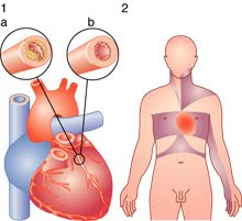
Angina pectoris is the medical term for chest pain or discomfort due to coronary heart disease. Angina is a symptom of a condition called myocardial ischemia. It occurs when the heart muscle (myocardium) doesn't get as much blood (hence as much oxygen) as it needs. This usually happens because one or more of the heart's arteries (coronary blood vessels that supply blood to the heart muscle) is narrowed or blocked. Insufficient blood supply is called ischemia.

The term derives from the Latin angina ("infection of the throat") from the Greek γχόνη ankhone ("strangling"), and the Latin pectus ("chest"), and can therefore be translated as "a strangling feeling in the chest".

Angina also can occur in people with valvular heart disease, hypertrophic cardiomyopathy (this is an enlarged heart due to disease) or uncontrolled high blood pressure. These cases are rare, though.

Typical angina is uncomfortable pressure, fullness, squeezing or pain in the center of the chest. The discomfort also may be felt in the neck, jaw, shoulder, back or arm. Many types of chest discomfort aren't related to angina. Acid reflux (heartburn) and lung infection or inflammations is examples.

Development of Angina Pectoris:

Angina pectoris is typically experienced in the symptoms of uncomfortable pressure, which feels like squeezing or pain in the center of the chest. You may also feel it in the neck, jaw, shoulder, back, arm, or stomach, including nausea.

It has been described as a tight or heavy feeling in the chest, or sometimes as pressure, squeezing, or burning. It can feel very difficult to breath, as though you are short of breath and unable to fill your lungs completely. These symptoms can be accompanied by numbness or tingling in the shoulders, arms, or wrists. However, just because you have these symptoms does not mean you have angina. You could have pericarditis, the inflammation of the sac around the heart. It might be something very serious, such as an aortic dissection, in which the inner layers of the aorta separate, causing you pain.

It might be a coronary spasm, in which the blood vessels to the heart spasm and constrict the blood flow momentarily. You may even be having a heart attack, so these painful symptoms should be evaluated by your doctor immediately. Even some non-heart related conditions can cause angina, such as heartburn, panic attacks, pleurisy (an inflammation of the chest membrane), esophageal spasms (in which the esophagus spasms causing food to back up), or simple sort muscles from improper stretching, or from conditions such as fibromyalgia. You may also have a lung infection or inflammation.


Angina, the feeling of pain or pressure, occurs when the heart needs more blood to bring it oxygen, so strenuous activity might trigger it. Running or walking quickly, carrying groceries, a rush of strong emotions, lifting something heavy, eating too much, having sex, or even extremes in temperatures, might trigger an attack. In some people, angina is caused by a condition known as coronary artery spasm, and in those cases you might feel pain or pressure even when you are resting.


Classification of Angina Pectoris:

There are two major different types of angina pectoris. There’s stable angina, and in these cases, the angina is usually predictable and you can expect to have symptoms in consistent situations (every time you run, every time you get angry, etc). Unstable angina means that the pain takes you by surprise, and can occur when you are at rest. With unstable angina, the chest pain can be more severe and prolonged than with stable angina. Both of these conditions can be caused by reduced blood flow to the heart muscles because of blockage by fatty build-ups. This blockage is called arterioschlerosis.

Stable angina:

Also known as effort angina, this refers to the more common understanding of angina related to myocardial ischemia. Typical presentations of stable angina is that of chest discomfort and associated symptoms precipitated by some activity (running, walking, etc.) with minimal or non-existent symptoms at rest. Symptoms typically abate several minutes following cessation of precipitating activities and resume when activity resumes. In this way, stable angina may be thought of as being similar to claudication symptoms.

People with stable angina (or chronic stable angina) have episodes of chest discomfort that are usually predictable. They occur on exertion (such as running to catch a bus) or under mental or emotional stress. Normally the chest discomfort is relieved with rest, nitroglycerin or both.

People with episodes of chest discomfort should see their physician for an evaluation. The doctor will evaluate the person's medical history and risk factors, conduct a physical exam, order a chest X-ray and take an electrocardiogram (ECG). Some people will also need an exercise ECG (stress test), an echocardiogram or other tests to complete the diagnosis.

Unstable angina:

Unstable angina (UA) (also "crescendo angina;" this is a form of acute coronary syndrome) is defined as angina pectoris that changes or worsens.

It has at least one of these three features:

  1. it occurs at rest (or with minimal exertion), usually lasting >10 min;
  2. it is severe and of new onset (i.e., within the prior 4–6 weeks); and/or
  3. it occurs with a crescendo pattern (i.e., distinctly more severe, prolonged, or frequent than previously).

UA may occur unpredictably at rest which may be a serious indicator of an impending heart attack. What differentiates stable angina from unstable angina (other than symptoms) is the pathophysiology of the atherosclerosis. The pathophysiology of unstable angina is the reduction coronary flow due to transient platelet aggregation on apparently normal endothelium, coronary artery spasms or coronary thrombosis. The process starts with atherosclerosis, and when inflamed leads to an active plaque, which undergoes thrombosis and results in acute ischemia, which finally results in cell necrosis after calcium entry. Studies show that 64% of all unstable anginas occur between 10 PM and 8 AM when patients are at rest.

In people with unstable angina, the chest pain is unexpected and usually occurs while at rest. The discomfort may be more severe and prolonged than typical angina or be the first time a person has angina. The most common cause is reduced blood flow to the heart muscle because the coronary arteries are narrowed by fatty buildups (atherosclerosis). An artery may be abnormally constricted or partially blocked by a blood clot. Inflammation, infection and secondary causes also can lead to unstable angina. In a form of unstable angina called variant or Prinzmetal's angina, the cause is coronary artery spasm.

Major risk factors:

  • Age (≥ 55 yo for men, ≥ 65 for women)
  • Cigarette smoking
  • Diabetes mellitus (DM)
  • Dyslipidemia
  • Family History of premature CVD
  • Hypertension (HTN)
  • Kidney disease
  • Obesity (BMI ≥ 30 kg/m2)
  • Physical inactivity
  • Smoking

Other Medical Problems by Angina Pectoris:

  • profound anemia
  • uncontrolled HTN
  • hyperthyroidism
  • hypoxemia
  • tachyarrhythmia
  • bradyarrhythmia
  • valvular heart disease
  • hypertrophic cardiomyopathy


Prevention of Angina Pectoris:

Unlike some diseases, however, angina can be stopped before it even gets started. It is quite preventable, but even if you haven’t prevented it, you can reduce it through some basic lifestyle changes. First, stop smoking. Smokers have more than double the risk of heart attacks and strokes than non-smokers, and heavy smokers have more than twice the risk of light smokers. Adopt healthier eating habits; avoid trans fats, saturated fats, cholesterol, and salt. A healthier diet will also help you control your blood pressure and overall cholesterol levels. Another overlooked lifestyle change concerns stress; can you relax, even when life is challenging? Learn to breathe deeply to keep oxygen flowing, which is necessary both to your heart and your feeling of well-being.

While it’s true that making good lifestyle choices can go far in preventing the heart disease that angina signals, it’s also true that some people are just prone to it because of their family history. Make sure to tell your doctor if anyone in your family has had heart disease or a heart attack or stroke. This information will help your doctor decide when and how to treat your angina.

Signs and Symptoms:

Most patients with angina complain of chest discomfort rather than actual pain: the discomfort is usually described as a pressure, heaviness, tightness, squeezing, burning, or choking sensation. Apart from chest discomfort, anginal pains may also be experienced in the epigastrium (upper central abdomen), back, neck area, jaw, or shoulders. This is explained by the concept of referred pain, and is due to the spinal level that receives visceral sensation from the heart simultaneously receiving cutaneous sensation from parts of the skin specified by that spinal nerve's dermatome, without an ability to discriminate the two. Typical locations for referred pain are arms (often inner left arm), shoulders, and neck into the jaw. Angina is typically precipitated by exertion or emotional stress. It is exacerbated by having a full stomach and by cold temperatures. Pain may be accompanied by breathlessness, sweating and nausea in some cases. In this case, the pulse rate and the blood pressure increases. The pain usually lasts for about 3 to 5 minutes but the symptoms actually starts 15 to 20 minutes before the cardiac arrest and the pain is relieved by rest or specific anti-angina medication. Chest pain lasting only a few seconds is normally not angina.


Myocardial ischemia comes about when the myocardia (the heart muscles) receive insufficient blood and oxygen to function normally either because of increased oxygen demand by the myocardia or by decreased supply to the myocardia. This inadequate perfusion of blood and the resulting reduced delivery of oxygen and nutrients is directly correlated to blocked or narrowed blood vessels.

Treatment of Angina Pectoris:

The most specific medicine to treat angina is nitroglycerin. It is a potent vasodilator that makes more oxygen available to the heart muscle. Beta-blockers and calcium channel blockers act to decrease the heart's workload, and thus its requirement for oxygen. Treatments are baloon angioplasty, in which the balloon is inserted at the end of a catheter and inflated to widen the arterial lumen. Stents to maintain the arterial widening are often used at the same time. Coronary bypass surgery involves bypassing constricted arteries with venous grafts. This is much more invasive than angioplasty.

The main goals of treatment in angina pectoris are relief of symptoms, slowing progression of the disease, and reduction of future events, especially heart attacks and, of course, death. Beta blockers (e.g., carvedilol, propranolol, atenolol) have a large body of evidence in morbidity and mortality benefits (fewer symptoms, less disability and longer life) and short-acting nitroglycerin medications have been used since 1879 for symptomatic relief of angina. Calcium channel blockers (such as nifedipine (Adalat) and amlodipine), isosorbide mononitrate and nicorandil are vasodilators commonly used in chronic stable angina [citation needed]. A new therapeutic class, called If inhibitor, has recently been made available: ivabradine provides pure heart rate reduction leading to major anti-ischemic and antianginal efficacy. ACE inhibitors are also vasodilators with both symptomatic and prognostic benefit and, lastly, statins are the most frequently used lipid/cholesterol modifiers which probably also stabilize existing atheromatous plaque [citation needed]. Low-dose aspirin decreases the risk of heart attack in patients with chronic stable angina, and was previously part of standard treatment, however it has since been discovered that the increase in haemorrhagic stroke and gastrointestinal bleeding offsets this gain so they are no longer advised unless the risk of myocardial infarction is very high.

Bookmark and Share